Undulation of the Retina on OCT

Sanjay Sharma MD, MSc (Epid), MBA
Professor of Ophthalmology, Queen’s University
A 64 year-old presents with blurred vision.
This image is consistent with which diagnosis?
- Epiretinal membrane
- Choroidal folds
- Central serous retinopathy
- Choroidal nevus
Analysis
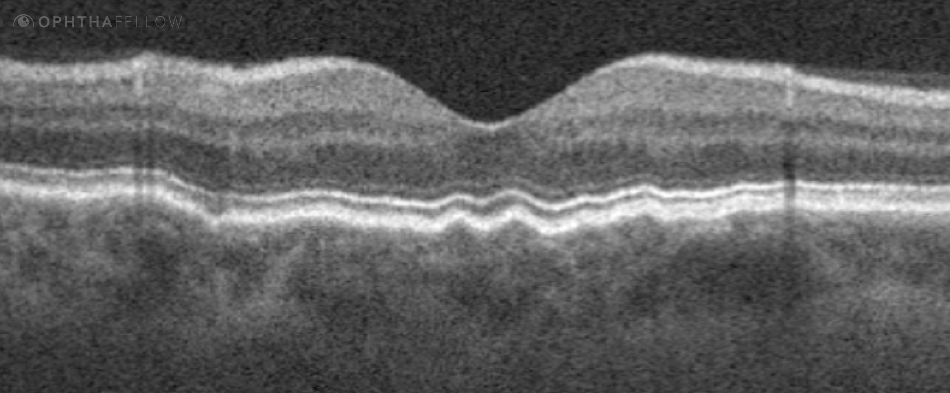
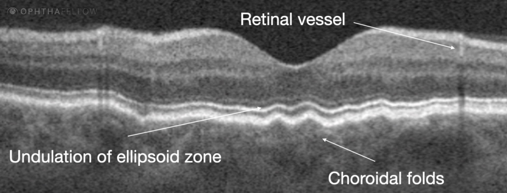
The inner and middle retina have normal lamellar architecture with clearly delineated NFL, GC, IPL, INL, OPL and ONL. There is undulation of the external limiting membrane, ellipsoid, and RPE
The patient was diagnosed with choroidal folds. Imaging confirmed the presence of an associated optic nerve meningioma.
The other options are less likely for the following reasons:
- An epiretinal membrane is usual visible as a thin hyperreflective line on the surface of the retina on OCT, which may be associated with superficial undulation, but is not associated with deeper folds.
- Central serous typically presents with sub retina fluid, a small focal pigment epithelial detachment with or without evidence of choroidal thickness. If the fluid is chronic, there may be elongation of the photoreceptors.
- Choroidal nevus may present with choriocapillaris hyperreflectivity and thickening of the choroid. Subretinal hyperreflective material or sub retinal fluid warrants suspicion for an associated choroidal neovascular membrane.
Video Analysis
In this tip’s accompanying 15 min. video, we will review:
Clinical Tip
In any patient who presents with choroidal folds, consider the possibility of autoimmune, thyroid or compressive lesion.