White Lesion on Cornea

Beeran Meghpara MD
Co-Director of the Refractive Surgery Department
Wills Eye Hospital, Philadelphia
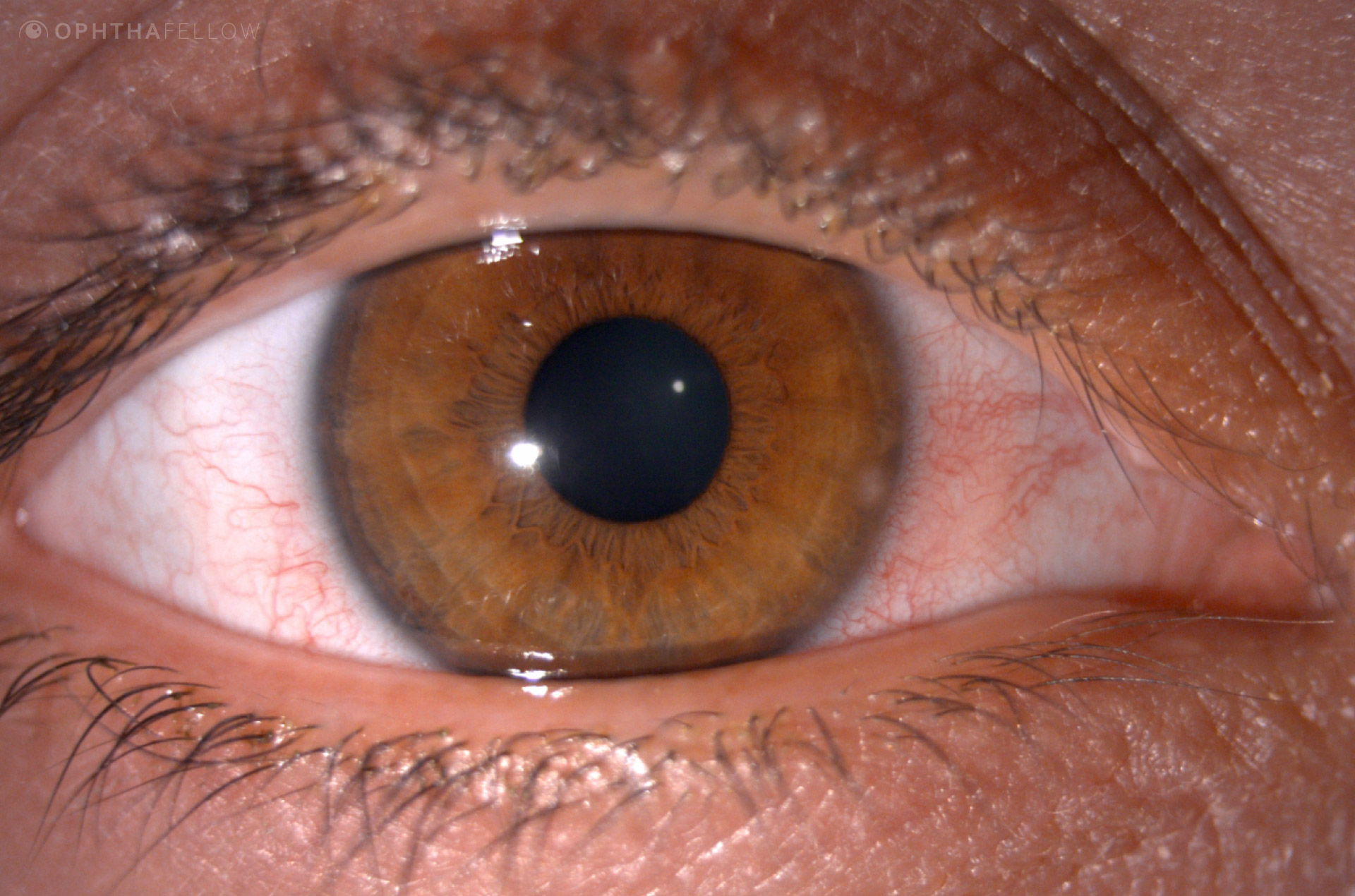
A 31-year-old presents with a 3-day history of pain and photophobia. Vision is 20/30. Why?
This image is most consistent with which diagnosis?
- Horner’s syndrome
- Peripheral corneal ulcer
- Granulomatous uveitis
- Herpes simplex keratitis
Analysis
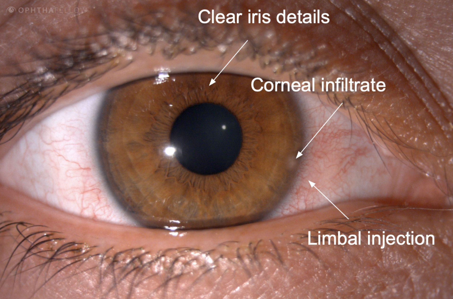
The patient was noted to have perilimbal injection and a mild increase in conjunctival vascularity. A small corneal infiltrate is seen at the 3:30 position on the peripheral cornea. There is no overlying epithelial defect and the iris and anterior chamber is clear.
The patient was diagnosed with a small peripheral corneal ulcer suggestive of a staphylococcal infection.
Video Analysis
In this 9-minute video featuring Dr. Beeran Meghpara, co-director of the Refractive Surgery Department at Wills Eye Hospital, you will:
Clinical Tip
In a small peripheral ulcer in which the epithelium is intact corneal cultures are not necessary and non-fortified topical antibiotics should be given.